
Advanced Dermatologic Surgery
Epidermoid Cyst Removal in Kansas City
Dr. Hocker is a triple board-certified dermatologist, dermatopathologist, and Mohs micrographic surgeon at Advanced Dermatologic Surgery, a national referral center in Overland Park, Kansas for melanoma, high-risk skin cancer, advanced reconstructive plastic surgery, and advanced tissue diagnostics. Dr. Hocker's triple board certification allows ADS to integrate surgical removal, refined reconstruction, and advanced laboratory studies on patients' tissue when clinically indicated. ADS also provides discreet surgical care for executives, public-facing professionals, Fortune 500 leaders, and other high-profile patients across the Midwest who value privacy, scar quality, and same-day reconstruction.
Epidermoid Cyst Removal: The Practical Summary
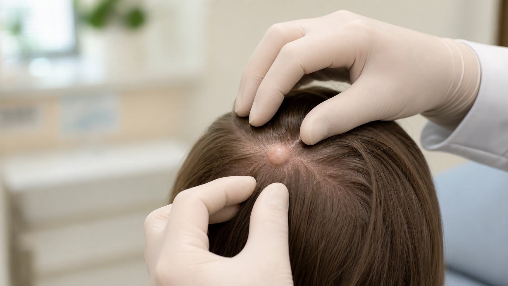
An epidermoid cyst is a benign keratin-filled sac under the skin, often with a small central pore called a punctum. It can be observed when it is small and quiet, but removal is usually best when it grows, hurts, drains, ruptures, recurs, sits in a visible area, or needs pathology to confirm the diagnosis.
What to remember
The cyst wall is the engine. Drainage empties the cyst, but complete excision removes the structure that keeps making keratin.
Key takeaways
- The central pore, or punctum, is the classic clue that separates many epidermoid cysts from lipomas and other lumps.
- Drainage can calm an inflamed cyst, but it usually does not remove the cyst wall, so recurrence is common.
- The lowest-recurrence surgery removes the entire cyst wall and includes the punctum when one is present.
- Inflamed cysts are often ruptured cysts, not true bacterial infections; antibiotics are added when cellulitis is present.
- Excised cysts should be submitted for permanent-section pathology when diagnostic certainty matters.
Evidence snapshot
- Epidermoid cysts are common benign cutaneous growths, and surgical technique affects recurrence and scar quality ( Mehrabi et al., 2002).
- A randomized trial found shorter wounds and faster operative times with punch incision compared with elliptical excision, without a statistically significant recurrence difference ( Lee et al., 2006).
- Multiple epidermoid cysts in a young patient can occasionally be a clue to inherited syndromes such as familial adenomatous polyposis/Gardner syndrome ( Half et al., 2009).
- In a review of inflamed epidermal inclusion cysts, 47% of cultures from mildly inflamed cysts showed no bacterial growth or only normal flora, supporting careful antibiotic use ( Meister et al., 2021).
What is an epidermoid cyst?
An epidermoid cyst is a non-cancerous skin growth that forms when keratin-producing surface skin cells become trapped under the skin.
The result is a smooth, round, slow-growing lump that often has a visible opening at the surface. Patients often call these “sebaceous cysts,” but that name is usually inaccurate. Epidermoid cysts contain keratin, not oil from sebaceous glands.
They most often occur on the face, neck, scalp, chest, shoulders, and back. They can appear almost anywhere, and they may stay quiet for years before becoming inflamed, tender, draining, or bothersome.
What causes epidermoid cysts to form?
Epidermoid cysts form when epidermal cells move into the dermis and keep producing keratin inside a walled-off sac.
This can happen around a blocked follicle, after acne, after skin trauma, or around an old scar. Most cysts are sporadic and benign.
Multiple epidermoid cysts in a child or young adult deserve more attention, especially with osteomas, desmoid tumors, dental abnormalities, or a family history of colon cancer. In that setting, Gardner syndrome, a variant of familial adenomatous polyposis related to APC gene mutations, may need evaluation ( Half et al., 2009).
When should an epidermoid cyst be removed?
A quiet, stable epidermoid cyst does not always need treatment. Removal is recommended when the cyst is changing, symptomatic, cosmetically important, repeatedly inflamed, or diagnostically uncertain.
| Reason | Why it matters |
|---|---|
| Growth | Growth raises the chance of rupture, inflammation, and diagnostic uncertainty. |
| Pain or drainage | Pain, odor, or drainage often means the cyst wall has ruptured or become irritated. |
| Visible location | A planned scar is usually better than an inflammatory scar after rupture. |
| Repeated flares | Repeated inflammation usually means the cyst wall remains and keeps refilling. |
| Uncertain diagnosis | Pilomatricoma, dermoid cyst, adnexal tumors, and rare cancers can mimic a cyst. |
How is an inflamed or infected cyst treated?
An inflamed cyst is often a ruptured cyst wall with keratin spilling into the surrounding skin, not a true bacterial infection.
That sterile inflammatory reaction can look infected even when the main problem is rupture. The right sequence is usually to calm the flare first, then remove the cyst later when the tissue planes are clearer.
| Cyst status | Best first step | Excision timing |
|---|---|---|
| Quiet and stable | Elective complete excision if desired | Same day or scheduled |
| Inflamed and painful | Drainage or steroid when appropriate | After several quiet weeks |
| Cellulitis present | Drainage plus antibiotics when clinically indicated | After infection resolves |
| Repeatedly inflamed | Control flare, then plan definitive removal | Often 4 to 8 weeks later |
Sources: Zuber, 2002; Stevens et al., 2014; Meister et al., 2021.
A surgeon's perspective: In my practice, the question is not just "Can I remove this today?" It is "Will removing it today give this patient the best operation?" A quiet cyst gives me clean planes. A ruptured, angry cyst often needs a calmer first step and a better-timed excision.
For primary care clinicians, the practical point is antibiotic stewardship. IDSA guidance does not recommend routine Gram stain and culture of pus from inflamed epidermoid cysts, and incision and drainage is the recommended first procedure when drainage is needed ( Stevens et al., 2014). Antibiotics are reserved for patients whose exam suggests surrounding cellulitis, systemic infection, immunosuppression, or another higher-risk situation.
Intralesional corticosteroid injection, commonly triamcinolone, can also be useful for selected acutely inflamed cysts when the goal is to calm inflammation before definitive excision. Some surgeons may offer single-stage excision for selected inflamed cysts, but when inflammation obscures the tissue planes, staged management often produces a cleaner operation.
How is an epidermoid cyst different from a lipoma, pilar cyst, or dermatofibroma?
Patients often use one phrase, such as “bump under the skin,” for several different lesions. The differences matter because each lesion has a different surgical plan, recurrence risk, and reason for pathology.
| Feature | Epidermoid cyst | Lipoma | Pilar cyst | Dermatofibroma |
|---|---|---|---|---|
| Texture | Firm or doughy | Soft, rubbery | Firm, dense | Firm, button-like |
| Central pore | Often present | Absent | Usually absent | Absent |
| Common site | Face, neck, back | Back, shoulders, arms | Scalp | Lower legs |
| Best clue | Punctum | Slides under finger | Scalp and family pattern | Dimple sign |
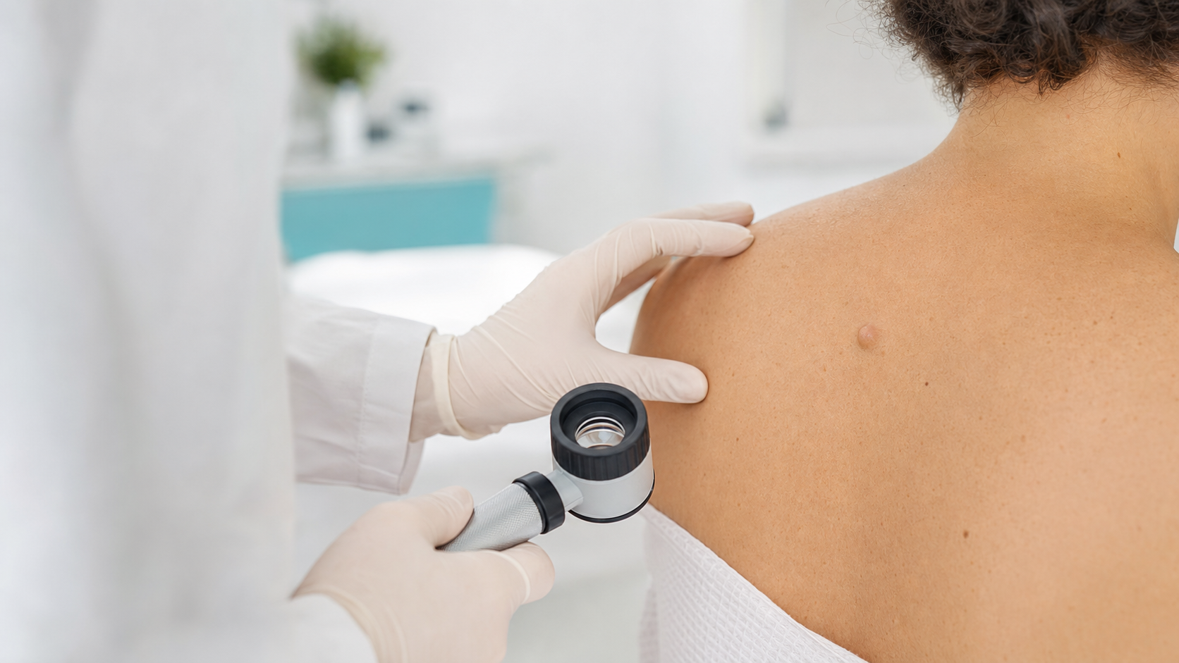
When the diagnosis is uncertain, ultrasound can be useful. It is especially helpful for deeper lumps, lesions that do not move like a typical cyst, or masses where the question is “skin lesion” versus “soft tissue mass” ( Achar et al., 2022).
What happens during complete cyst excision?
Complete cyst excision is an office procedure under local anesthesia. The goal is to remove the intact cyst wall because the wall is what keeps producing keratin.
Step 1: Local anesthesia
Step 2: Incision planning
Step 3: Cyst wall dissection
Step 4: Layered closure
Step 5: Pathology review
A surgeon's perspective: My bias is simple: remove the wall, not just the contents. If I only empty the white keratin, I have helped for the day. If I remove the wall and punctum, I have treated the problem.
What does recovery from epidermoid cyst excision look like?
Most patients return to desk work the same or next day. The scar continues to mature for 6–12 months.
Patients usually have mild soreness, swelling, and bruising for several days. Sutures are often removed around 7 days on the face and 10–14 days on the body, though timing varies by location and closure type.
Scar quality depends on incision planning, body location, wound tension, and aftercare. The chest, shoulders, and back tend to form wider scars than the face because the skin is under more tension. Sun protection, scar massage when appropriate, and silicone gel or silicone sheets can improve scar appearance over time.
Can an epidermoid cyst become cancer?
Epidermoid cysts are benign, and cancer arising inside one is rare. Published estimates for squamous cell carcinoma arising within an epidermoid cyst are roughly 0.011%–0.045%.
That low number should not cause panic. It should create a sensible rule: a routine, stable cyst can often be watched, but a changing cyst deserves attention.
Squamous cell carcinoma is the malignancy most often discussed in this setting, but basal cell carcinoma, Bowen disease, melanoma in situ, and other tumors have also been reported within or adjacent to cyst-like lesions in case literature ( Shen-Wagner et al., 2024). The teaching point is not that ordinary cysts are dangerous. The teaching point is that a lesion that stops behaving like a routine cyst should not be treated casually.
Which cyst changes should prompt evaluation?
How does Advanced Dermatologic Surgery approach cyst removal?
At Advanced Dermatologic Surgery, Dr. Hocker approaches cyst removal as diagnosis-first dermatologic surgery: confirm what the lesion is, remove it with the right technique, and plan the scar before the first incision.
Most routine epidermoid cysts do not require a cancer-center-level surgeon. The value of a surgical dermatologist is highest when the cyst is on the face, ear, scalp, eyelid, lip, or neck; when it is recurrent or inflamed; when scar quality matters; or when the diagnosis is not completely routine.
Dr. Hocker’s triple board certification in dermatology, dermatopathology, and Mohs surgery is most relevant when a lesion behaves atypically, sits in a high-visibility area, or requires real-time surgical judgment. Advanced Dermatologic Surgery has an on-site CLIA-certified pathology laboratory used for Mohs surgery and intraoperative frozen-section work when medically necessary. Routine benign excision specimens such as typical epidermoid cysts, pilar cysts, lipomas, and dermatofibromas are not the usual purpose of the in-office lab. But if an operation reveals something unusual, Dr. Hocker’s dermatopathology training and the availability of on-site frozen-section capability allow real-time diagnostic escalation when needed.
A surgeon's perspective: My bias is to make the operation fit the lesion. A small, quiet back cyst can often be handled simply. A recurrent cyst on the face, an inflamed cyst with distorted tissue planes, or a "cyst" that feels atypical deserves more deliberate planning.
Advanced Dermatologic Surgery is located at 6901 W 121st Street in Overland Park, Kansas, serving Kansas City, Leawood, Olathe, Lenexa, Prairie Village, and surrounding communities.
Frequently Asked Questions
Should I pop an epidermoid cyst at home?
Will an epidermoid cyst come back after removal?
Are epidermoid cysts cancerous?
How much does epidermoid cyst removal cost?
Where can I have an epidermoid cyst removed in Kansas City?
Do I need to stop blood thinners before cyst removal?
How long is recovery after cyst removal?
Does every epidermoid cyst need pathology?
When should a primary care clinician refer a cyst?
Can ultrasound help with a lump under the skin?
What if the cyst looks unusual during surgery?
When should you schedule an evaluation?
Schedule an evaluation if a cyst is new, growing, painful, draining, changing, repeatedly inflamed, cosmetically important, or diagnostically uncertain.
Schedule a consultation