

Advanced Dermatologic Surgery
Pilar Cyst Removal in Kansas City
Dr. Hocker is a triple board-certified dermatologist, dermatopathologist, and Mohs micrographic surgeon at Advanced Dermatologic Surgery, a national referral center in Overland Park, Kansas for melanoma, high-risk skin cancer, advanced reconstructive plastic surgery, and advanced tissue diagnostics. For scalp cyst surgery, that background matters most in diagnosis-focused surgical judgment: deciding whether a cyst behaves routinely, when pathology matters, and how to protect the hair-bearing scalp.
Pilar Cyst Removal: The Practical Summary
A pilar cyst, also called a trichilemmal cyst, is a firm keratin-filled cyst that usually forms on the scalp. Most are benign, slow-growing, and removable in the office under local anesthesia. Removal is most useful when the cyst is growing, painful, catching on combs or hats, repeatedly inflamed, cosmetically bothersome, or not behaving like an ordinary cyst.
What to remember
The most important decision is not whether a scalp lump has been called a "cyst." The important decision is whether it behaves like a routine pilar cyst: slow-growing, smooth, mobile, and intact. A cyst that grows rapidly, ulcerates, bleeds, becomes fixed, recurs after prior surgery, or reaches a large size deserves a more careful plan and pathology review.
Key takeaways
- Pilar cysts are usually benign scalp cysts filled with compact keratin.
- They often feel firmer and smoother than epidermoid cysts and usually lack a central pore.
- Complete removal of the cyst wall is what lowers recurrence risk.
- Inflamed or ruptured cysts are harder to remove cleanly; timing matters.
- A small minority can develop proliferating pilar tumor changes, so unusual clinical features should not be ignored.
Evidence snapshot
- Classic hereditary pilar cyst literature describes affected families, scalp predominance, encapsulated cysts, and autosomal dominant inheritance in many families ( Leppard et al., 1977).
- PLCD1 variants are linked to hereditary trichilemmal cyst formation, though routine genetic testing is not needed for most patients ( Horer et al., 2019; Kolodney et al., 2020).
- DermNet describes trichilemmal cysts as scalp-predominant, firm, mobile nodules that usually lack a central punctum ( DermNet, 2014).
- Review literature reports that up to about 2% of trichilemmal cysts can give rise to proliferating pilar tumors ( Alshaalan et al., 2021).
- A 2024 systematic review found 361 published proliferating pilar tumor cases, with 36.8% classified as malignant; this is not the cancer risk of an ordinary pilar cyst ( Nemeh et al., 2024).
- Malignant trichilemmal cysts account for less than 0.1% of all skin cancers, which is reassuring for patients but still supports careful evaluation of rapidly changing lesions ( Shen-Wagner et al., 2024).
- In a punch-incision series of keratinous and pilar cysts, recurrence was 3.6% by chart review and 8.3% among survey responders ( Mehrabi et al., 2002).
What is a pilar cyst?
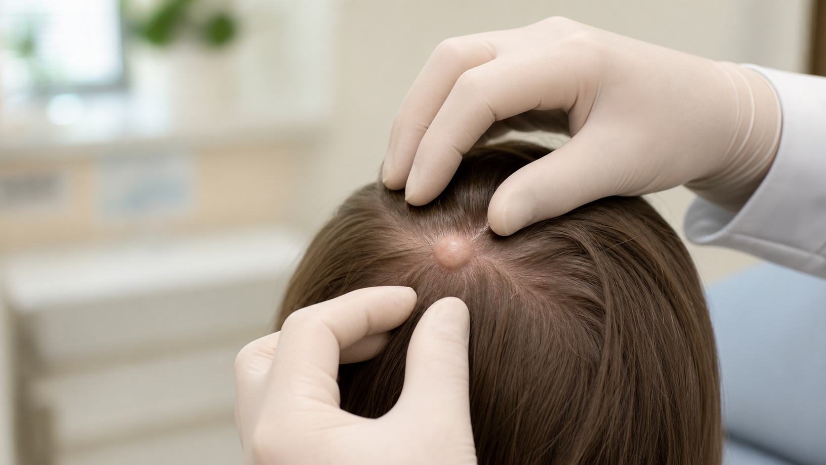
A pilar cyst is a benign cyst that forms from the outer root sheath of a hair follicle. It contains keratin, the same structural protein found in hair and nails, but the keratin is packed into a firm cyst rather than released normally through the skin.
Most patients notice a pilar cyst as a smooth bump under the scalp. It may move slightly under the skin. It may be painless for years. It may also become tender if it is irritated by combs, hats, headphones, helmets, or repeated pressure.
Pilar cysts are often mistakenly called "sebaceous cysts." That name is common, but it is not very precise. A pilar cyst is not a blocked oil gland. It is a keratin-filled follicular cyst with a wall that behaves differently from an epidermoid cyst.
How is a pilar cyst different from an epidermoid cyst?
A pilar cyst usually occurs on the scalp, feels firm and smooth, and has no central pore. An epidermoid cyst more often has a punctum, is more attached to the skin surface, and may rupture more easily.
| Feature | Pilar cyst | Epidermoid cyst |
|---|---|---|
| Common location | Scalp | Face, neck, trunk, back, groin, many sites |
| Surface clue | Usually no punctum | Often has a central punctum |
| Texture | Firm, smooth, dense, sometimes doughy | Firm to fluctuant; may feel softer if inflamed |
| Contents | Compact keratin | Softer keratin debris |
| Wall behavior | Often thicker and more durable | Often thinner and more rupture-prone |
| Histopathology | Trichilemmal keratinization, classically abrupt keratinization without a granular layer | Epidermal-type keratinization with a granular layer |
| Surgical goal | Remove intact cyst wall and contents | Remove entire wall after inflammation is controlled |
The distinction matters because the surgery is different. A calm pilar cyst on the scalp often shells out cleanly. A ruptured or inflamed cyst behaves more like a sticky problem: the normal tissue planes are distorted, and complete wall removal becomes harder.
Are pilar cysts hereditary?
Yes, some pilar cysts are hereditary. The classic pattern is autosomal dominant inheritance with incomplete penetrance, meaning a parent can pass along the tendency even though not every person who inherits it develops obvious cysts.
Patients often tell a very recognizable story: "My mother had these," "My grandmother had scalp cysts," or "Several women in my family have had them removed." That pattern is real. Leppard and colleagues described hereditary pilar cysts in families in 1977, and later genetic studies connected hereditary trichilemmal cysts to PLCD1 variants ( Leppard et al., 1977; Horer et al., 2019; Kolodney et al., 2020).
For most patients, that genetic detail does not change the immediate plan. It mainly explains why one person may develop several scalp cysts over time and why new cysts can appear even after a prior cyst was removed completely.
Can a pilar cyst become cancerous?
An ordinary pilar cyst is usually benign. The concern is not that every scalp cyst is dangerous. The concern is that a long-standing or changing pilar cyst can rarely develop into a proliferating pilar tumor, and an even smaller subset of those tumors can behave malignantly.
Patients should not be frightened into thinking every scalp bump is cancer. But patients and primary care clinicians should recognize the warning signs that move a cyst out of the routine category.
| Feature | Usually routine | Needs more urgent evaluation |
|---|---|---|
| Growth speed | Slow change over years | Rapid growth over weeks or months |
| Skin surface | Normal scalp skin | Ulceration, bleeding, crusting, drainage |
| Feel | Smooth, mobile, round | Fixed, irregular, firm, rock-like, tender |
| Size | Small to moderate and stable | Large, especially greater than 5 cm or expanding |
| History | New or stable cyst | Recurrence after previous removal |
Sources: Alshaalan et al., 2021; Nemeh et al., 2024; Ye et al., 2004.
Published reviews report that up to about 2% of trichilemmal cysts may give rise to proliferating trichilemmal tumors ( Alshaalan et al., 2021). A 2024 systematic review found 361 published proliferating pilar tumor cases, with 36.8% classified as malignant ( Nemeh et al., 2024). That 36.8% number is not the cancer risk of a normal pilar cyst. It is the proportion of reported proliferating pilar tumor cases that were classified as malignant in the published literature. Another useful patient-facing anchor is that malignant trichilemmal cysts account for less than 0.1% of all skin cancers ( Shen-Wagner et al., 2024).
For practical purposes, remember this: a quiet, stable scalp cyst is usually a benign surgical problem. A rapidly growing, ulcerated, bleeding, recurrent, fixed, or unusually large scalp tumor needs a diagnosis-focused plan.
For referring providers: not every "cyst" is a cyst
Most scalp cysts are benign, but clinical-pathologic mismatch is common enough that changing, recurrent, ulcerated, bleeding, fixed, unusually large, or clinically odd lesions should be treated as diagnostic problems.
- In a 2,438-case clinicopathologic series of cutaneous cysts, trichilemmal cysts were the second most common cutaneous cyst overall at 23.8%, and clinical-pathologic mismatch was common enough to support histologic confirmation when lesions are removed ( Kamyab et al., 2020).
- In an adult scalp-mass surgical series, trichilemmal cyst was the most common diagnosis, but clinically significant pathology was found in about 7.8% of cases and preoperative diagnostic accuracy across departments was low ( Turk et al., 2015).
- In a 2024 review of lesions with "cyst" in the clinical differential, nearly 1 in 20 were malignant on histopathology, most commonly basal cell carcinoma or squamous cell carcinoma ( McCrary et al., 2024).
That does not mean every scalp cyst needs urgent removal. It means the referral question should be: does this lesion behave like an ordinary pilar cyst, or does it need diagnosis-focused excision and pathology?
When should a pilar cyst be removed?
A pilar cyst should be removed when it is symptomatic, growing, repeatedly inflamed, catching on grooming or headwear, visible through thinning hair, diagnostically uncertain, or personally bothersome enough that the patient wants a definitive diagnosis.
Observation is reasonable when the cyst is small, stable, painless, and cosmetically unimportant. Removal becomes more compelling when the cyst interferes with daily life or stops behaving predictably.
It catches on combs, hats, helmets, headphones, or pillows
It hurts or becomes tender with pressure
It is growing or changing
It is visible through thinning hair
The diagnosis is uncertain
A surgeon's perspective: In my practice, the best time to remove a scalp cyst is when it is calm, intact, and still has clean tissue planes. A quiet pilar cyst often shells out beautifully. A ruptured, angry cyst turns a neat operation into a less predictable one.
What happens during pilar cyst excision?
Pilar cyst excision is usually performed in the office under local anesthesia. The surgeon numbs the scalp, makes a small planned incision, separates the cyst wall from surrounding tissue, removes the cyst, controls bleeding, and closes the incision so the scar hides as well as possible within the hair.
| Step | Why it matters |
|---|---|
| Hair parting and marking | Keeps the incision aligned with the cyst and scalp anatomy. |
| Local anesthesia | Allows the procedure to be done comfortably in the office. |
| Small planned incision | Gives access while respecting scar visibility and hair direction. |
| Careful dissection | Frees the cyst wall without shredding it. |
| Complete cyst removal | Lowers the chance of recurrence from retained wall. |
| Hemostasis | Matters on the scalp because it has a rich blood supply. |
| Precise closure | Helps narrow the final scar and protect nearby hair follicles. |
The thick wall of a pilar cyst can be an advantage. When it is intact, it often allows the cyst to be delivered cleanly. That is one reason patients should avoid squeezing or puncturing scalp cysts at home.
Published recurrence data are imperfect because techniques, cyst types, and follow-up methods vary. In one punch-incision series of keratinous and pilar cysts, recurrence was 3.6% by chart review and 8.3% among survey responders ( Mehrabi et al., 2002). The useful takeaway is simpler than the decimal: recurrence is generally low when the wall is removed completely, and it rises when cyst wall is left behind.
Should an infected or inflamed pilar cyst be removed right away?
Usually, no. An actively inflamed or infected cyst is often better treated first, then removed later after the inflammation settles.
Inflammation changes the operation. The cyst wall becomes more fragile. The surrounding tissue becomes swollen and sticky. The chance of incomplete wall removal rises. That is why the cleanest plan is often staged: calm the cyst first, then remove it once the planes return.
Depending on the exam, treatment may include incision and drainage, culture if bacterial infection is suspected, antibiotics when there is surrounding cellulitis, or a short anti-inflammatory approach. Definitive excision is often planned several weeks later, once the scalp has quieted down.
This is not delay for delay's sake. It is timing the operation so the final result is better. There is evolving evidence that selected inflamed epidermal cysts can sometimes be excised in a single stage by experienced surgeons, but that evidence is not specific to pilar cysts and does not erase the basic surgical principle: the calmer and more intact the cyst, the cleaner the operation is likely to be ( Gwak et al., 2026).
What is recovery like after scalp cyst removal?
Recovery after pilar cyst removal is usually straightforward. Most patients have mild soreness, scalp tightness, and modest swelling for several days. Sutures are commonly removed in about 10 to 14 days, depending on the closure and location.
Most desk work can often resume the same day or next day. Strenuous exercise is commonly limited for about one to two weeks, depending on bleeding risk and closure tension. Call promptly for spreading redness, warmth, worsening pain, fever, pus, sudden swelling, or bleeding that does not stop with steady pressure.
Scalp wounds often heal well because of strong blood supply. That same blood supply also means bleeding control and pressure dressing technique matter.
Dermatologic surgery complication rates are generally low, but the scalp deserves respect. In a prospective dermatologic surgery study of 1,911 patients, infection risk was 1.3% overall but greater than 3% on the scalp, and hemorrhage risk was 0.89% overall ( Bordeaux et al., 2011). That is why scalp cyst instructions focus on pressure, activity limits, and calling promptly for swelling, bleeding, spreading redness, or worsening pain.
Will hair grow back over the scar?
Usually, the surrounding hair hides the incision very well, but hair does not grow directly through scar tissue. The goal is a narrow, well-oriented scar that is concealed by nearby hair rather than a widened line that becomes visible.
This is especially important for patients with thinning hair, short hairstyles, public-facing work, or multiple cysts that may require staged removal. A pilar cyst may be benign, but scalp scar planning is not trivial.
A surgeon's perspective: My bias on the scalp is to plan the scar before I plan the incision length. The shortest incision is not always the best incision. The best incision is the one that removes the cyst cleanly and leaves the least visible evidence that surgery happened.
How does Advanced Dermatologic Surgery approach pilar cyst removal?
Advanced Dermatologic Surgery approaches pilar cyst removal as diagnosis-focused scalp surgery, not as a simple "bump removal." The goal is to confirm what the lesion is, remove appropriate cysts cleanly, protect the hair-bearing scalp, and escalate when a cyst behaves unusually.
For routine benign-appearing pilar cysts, the key surgical issues are incision planning, intact wall removal, bleeding control, closure, scar width, and recurrence prevention. For atypical cysts, the key issue is diagnostic safety: do not casually treat a rapidly growing, ulcerated, recurrent, fixed, or unusually large scalp tumor as an ordinary cyst.
Advanced Dermatologic Surgery has an on-site CLIA-certified pathology laboratory used for Mohs surgery and intraoperative frozen-section work when medically necessary. Routine benign pilar cyst specimens are not the usual purpose of the in-office lab. If a procedure reveals something unusual, Dr. Hocker's dermatopathology training and the availability of on-site frozen-section capability allow real-time diagnostic escalation when needed.
Advanced Dermatologic Surgery is located at 6901 W 121st Street, Overland Park, KS 66209, serving Overland Park, Kansas City, Leawood, Olathe, Lenexa, Prairie Village, and the broader Kansas City metro. Patients can call (913) 661-1755 or request an appointment online.
Frequently Asked Questions
Can I pop or drain a pilar cyst at home?
Do I need to shave my head for pilar cyst surgery?
Should all of my pilar cysts be removed if I have several?
Will a pilar cyst come back after removal?
Does insurance cover pilar cyst removal?
Do I need to stop blood thinners before scalp cyst removal?
How quickly will I get pathology results?
When should I worry about a scalp cyst?
When should you schedule an evaluation?
Schedule an evaluation if a scalp cyst is growing, painful, inflamed, catching on grooming, visible through hair, recurrent after previous removal, bleeding, ulcerated, larger than expected, or simply worrying you.
A good consultation should answer three questions: What is it? Does it need to come out? And if it does, what is the cleanest way to remove it while protecting the scalp and scar?
Schedule a consultation