
Advanced Dermatologic Surgery
Lipoma Removal in Kansas City
Dr. Hocker is a triple board-certified dermatologist, dermatopathologist, and Mohs micrographic surgeon at Advanced Dermatologic Surgery, a national referral center in Overland Park, Kansas for melanoma, high-risk skin cancer, advanced reconstructive plastic surgery, and advanced tissue diagnostics. Dr. Hocker's triple board certification allows ADS to integrate surgical removal, refined reconstruction, and advanced laboratory studies on patients' tissue when clinically indicated. ADS also provides discreet surgical care for executives, public-facing professionals, Fortune 500 leaders, and other high-profile patients across the Midwest who value privacy, scar quality, and same-day reconstruction.
Lipoma Removal: The Practical Summary
A lipoma is a benign growth of mature fat cells that usually feels soft, rubbery, and movable under normal skin. Most small, stable lipomas are harmless. Removal is reasonable when the lump grows, hurts, presses on a nerve, changes body contour, rubs on clothing, or has warning signs that require imaging or pathology.
What to remember
The practical danger is not that an ordinary lipoma commonly turns into cancer. The danger is assuming every fatty lump is ordinary. A large, deep, firm, painful, fixed, or rapidly growing fatty mass deserves a different level of evaluation.
Key takeaways
- Most lipomas are benign. A typical lipoma is soft, slow-growing, movable, and covered by normal skin.
- The exam changes the plan. Size, depth, texture, fixation, pain, and growth speed matter more than the word "lipoma."
- Five centimeters is a useful threshold. Masses at or above 5 cm, or deep to the fascia, deserve more caution and often imaging.
- Excision gives the cleanest answer when diagnosis matters because it preserves tissue for pathology.
- Liposuction-assisted removal is selective. It can reduce scar length in selected larger low-risk lipomas, but it may fragment tissue.
Evidence snapshot
- Clinically oriented reviews describe lipomas as approximately one-half of benign soft-tissue masses, while referral-center series use different denominators ( Achar et al., 2022 ; Murphey et al., 2004 ).
- High-risk mass features include size 5 cm or larger, deep location, rapid growth, or fixation ( Achar et al., 2022 ; Johnson et al., 2018 ).
- In a dermatologic surgery series, 8 of 637 lesions diagnosed before surgery as lipomas - 1.3% - were well-differentiated liposarcomas on pathology ( Sato et al., 2018 ).
- Complete excision has a low recurrence rate when the lesion is removed completely; one extremity series reported 0% recurrence for lipomas after marginal excision ( Errani et al., 2016 ).
What is a lipoma?
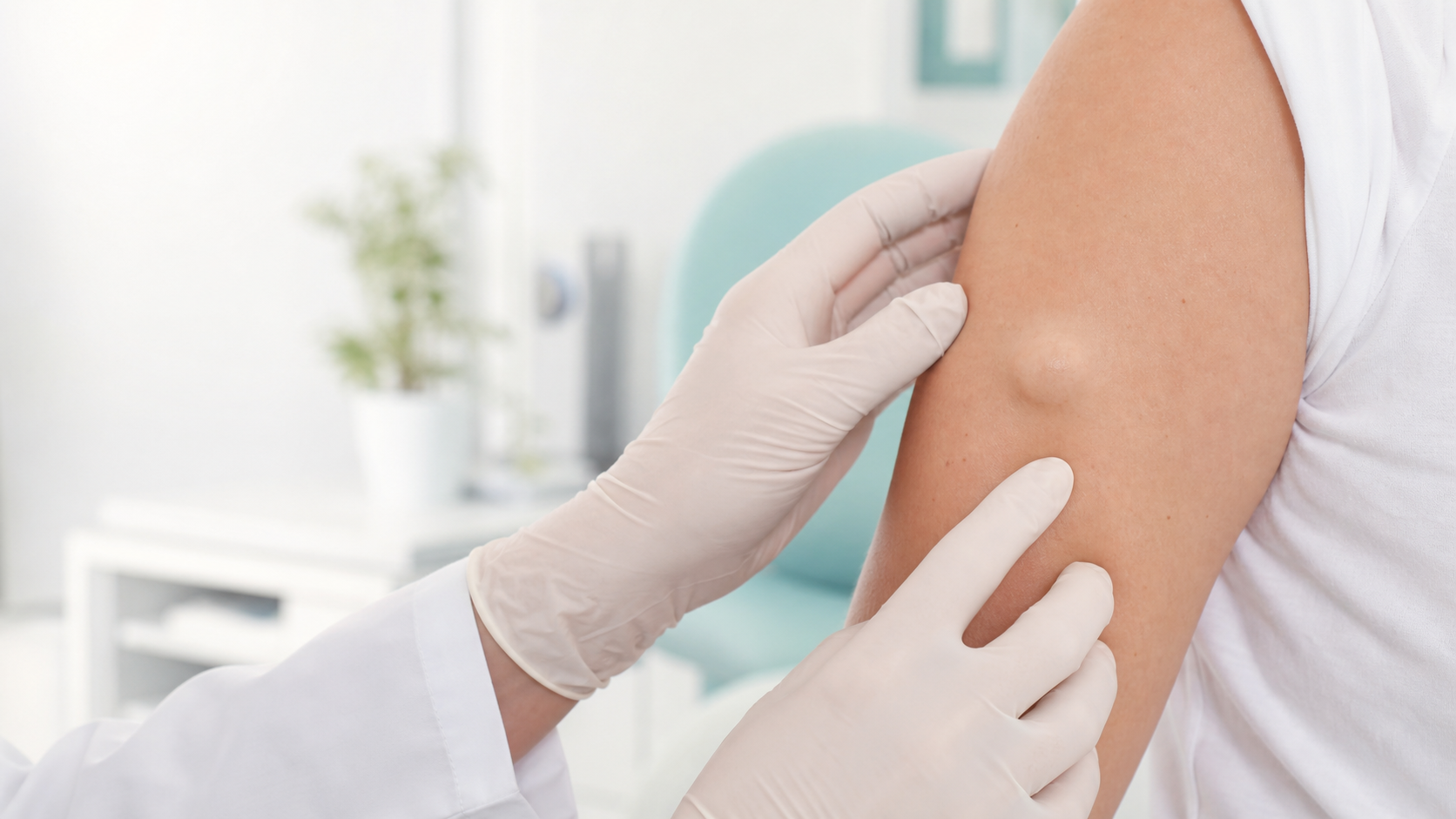
A lipoma is a benign tumor made of mature fat cells. It usually sits in the subcutaneous layer between the skin and the underlying muscle, where it feels soft, doughy, rubbery, and movable.
Patients often discover a lipoma by feel before they notice it visually. The classic story is simple: "I found a soft lump under my skin. It moves when I press on it. It has been there for months or years."
That story is reassuring, but it is not a diagnosis by itself. A good exam determines whether the lump is truly superficial, freely mobile, and soft, or whether it is deeper, firmer, fixed, painful, or growing faster than expected.
Lipomas commonly occur on the upper back, shoulders, neck, upper arms, forearms, abdomen, thighs, forehead, and scalp ( Salam, 2002 ). They are often noticed in adulthood, especially between ages 40 and 70 ( Achar et al., 2022 ).
How can you tell a lipoma from a cyst?
A lipoma usually feels soft and movable, with normal skin over it and no central pore. An epidermoid cyst is more likely to be dome-shaped, attached to the skin, and marked by a small central opening called a punctum.
Patients often call any lump a "cyst." That is understandable, but it matters surgically. A cyst has a wall and keratin contents. A lipoma is fatty tissue. The incision, dissection plane, recurrence risk, and pathology question are different.
| Feature | Lipoma | Epidermoid cyst | Lymph node | Liposarcoma concern |
|---|---|---|---|---|
| Texture | Soft, doughy | Firm or fluctuant | Rubbery or firm | Firm or hard |
| Mobility | Often very mobile | Moves with skin | Variable | Often fixed/deep |
| Skin surface | Usually normal | May have punctum | Normal | Usually normal |
| Contents | Mature fat | Keratin | Lymph tissue | Malignant fat tumor |
| Typical action | Observe or excise | Remove wall if needed | Evaluate cause | Image/refer/biopsy |
The key is not memorizing every lump. The key is knowing when the pattern does not fit.
When should a fatty lump be checked quickly?
A fatty lump should be checked promptly if it is larger than 5 cm, deep, rapidly growing, firm, fixed to deeper tissue, painful at rest, newly appearing without explanation, or located in a higher-risk area such as the thigh, retroperitoneum, or proximal extremity.
Most lipomas are harmless. Liposarcoma is rare. But soft-tissue cancers are serious enough that the warning signs deserve a clean, memorable rule.
| Warning sign | Why it matters |
|---|---|
| 5 cm or larger | Higher-risk size threshold. |
| Deep to fascia | Needs imaging clarity. |
| Rapid growth | Not typical lipoma behavior. |
| Firm or hard | Changes the diagnosis. |
| Fixed to muscle | Suggests deeper involvement. |
| Pain at rest | May signal nerve pressure or another diagnosis. |
Sources: Achar et al., 2022 ; Johnson et al., 2018.
A surgeon's perspective: In my practice, the safest question is not "Could this be a lipoma?" It is "Does this lump behave like a routine lipoma?" A soft, mobile, superficial lump that changes slowly is one category. A firm, deep, painful, fixed, fast-growing, or 5 cm lump is another.
Ultrasound is often a practical first imaging step because it can confirm whether a mass is solid or cystic and estimate size and depth. In a superficial soft-tissue tumor study, ultrasound identified lipomas with 95.2% sensitivity and 94.3% specificity ( Hung et al., 2014 ). MRI is more appropriate when a mass is deep to fascia, 5 cm or larger, enlarging rapidly, or otherwise atypical.
That does not mean every 5 cm lump is cancer. It means the evaluation should be more disciplined.
Can a lipoma turn into liposarcoma?
A typical superficial lipoma is not expected to routinely turn into liposarcoma. The more important clinical point is that a malignant fatty tumor can sometimes be mistaken for a benign lipoma before imaging or pathology clarifies the diagnosis.
That distinction matters because it keeps the message truthful without creating unnecessary fear.
Most soft, superficial, slow-growing, mobile fatty lumps are benign. But a fatty lump that is large, deep, firm, painful, fixed, or growing quickly should not be casually dismissed. Liposarcoma classically occurs in deeper locations such as the thigh and retroperitoneum and rarely arises from a typical superficial lipoma ( Springfield, 1993 ).
The miss rate is small but real. In a 10-year dermatologic surgery series, 8 of 637 lesions diagnosed before surgery as lipomas - 1.3% - were ultimately well-differentiated liposarcomas on pathology ( Sato et al., 2018 ). That is the right kind of number: reassuring for most patients, but strong enough to explain why atypical features should not be ignored.
For referring clinicians, one pathology detail is worth remembering: atypical lipomatous tumor/well-differentiated liposarcoma is often associated with MDM2 and CDK4 amplification. When imaging and histology are ambiguous, molecular testing such as MDM2 fluorescence in situ hybridization can help separate it from benign lipoma ( Yee et al., 2022 ). Patients do not need to remember the gene names. They need to remember the rule: an atypical fatty lump deserves an atypical workup.
The problem is not the soft, stable lipoma. The problem is the lump everyone kept calling a lipoma when it was never behaving like one.
When should a lipoma be removed?
A lipoma should be removed when it is growing, painful, pressing on a nerve, rubbing on clothing, changing a visible contour, functionally bothersome, diagnostically uncertain, or simply bothersome enough that the patient wants a definitive answer.
Observation is reasonable for a small, soft, stable lipoma that causes no symptoms and has no concerning features. Removal becomes reasonable when the lipoma affects comfort, function, appearance, or confidence in the diagnosis.
Growth or change
Pain or nerve symptoms
Visible contour change
Friction or pressure
Diagnostic uncertainty
What happens during lipoma removal?
Lipoma removal is usually an in-office procedure under local anesthesia. The surgeon plans an incision, numbs the area, opens the correct tissue plane, separates the lipoma from surrounding tissue, removes it as completely as possible, controls bleeding, and closes the wound in layers.
| Step | Why it matters |
|---|---|
| Marking | Preserves the plan after numbing. |
| Local anesthesia | Keeps the procedure comfortable. |
| Planned incision | Balances access and scar. |
| Capsule dissection | Separates lipoma from normal tissue. |
| Complete removal | Lowers recurrence risk. |
| Layered closure | Reduces dead space and tension. |
Small lipomas may take about 20 to 30 minutes. Larger, deeper, scar-sensitive, or anatomically complex lipomas may take longer. Recovery depends on size and location, but many patients return to desk work the same or next day.
Is excision better than liposuction for lipoma removal?
For most routine lipomas, standard excision is the most definitive option because it removes the mass directly and preserves tissue for pathology. Liposuction-assisted techniques can be useful in selected larger, low-risk lipomas when scar minimization is the dominant priority and the diagnosis is secure.
Plastic surgeons and dermatologic surgeons have both published reasonable approaches. The question is not "Which specialty is better?" The question is: what does this lump require?
| Factor | Standard excision | Liposuction-assisted |
|---|---|---|
| Best use | Most lipomas | Selected large, low-risk lipomas |
| Specimen | More intact | More fragmented |
| Capsule | Directly removed | May need adjunct removal |
| Scar | Linear | Smaller entry site |
| Diagnostic certainty | Stronger | Weaker if atypical |
Sources: Pinski & Roenigk, 1990 ; Choi et al., 2007 ; Copeland-Halperin et al., 2015.
A surgeon's perspective: My surgical bias is to protect the diagnosis before chasing the smallest possible incision. If a fatty lump is classic and low-risk, scar-sparing choices can be discussed. If it is atypical, the priority is a plan that gives a clean specimen and a reliable diagnosis.
What does recovery look like after lipoma removal?
Recovery is usually straightforward: soreness, bruising, and tightness for several days; suture removal based on location; exercise restrictions for one to two weeks; and scar maturation over six to twelve months.
The bigger the lipoma, the more important dead-space management becomes. After a fatty mass is removed, the body has to close down the pocket it occupied. That is why layered closure, pressure dressing, and temporary activity restriction matter.
Call the office promptly for spreading redness, increasing warmth, worsening pain, fever, drainage, sudden swelling, or bleeding that does not stop with pressure.
How does Advanced Dermatologic Surgery approach lipoma removal?
Advanced Dermatologic Surgery evaluates lipomas with a surgical dermatologist's eye: diagnose the lump first, decide whether imaging or removal is appropriate, plan the incision around both access and scar, and escalate diagnostically if the lump behaves unusually.
Dr. Thomas L.H. Hocker is triple board-certified in dermatology, dermatopathology, and Mohs micrographic surgery. For lipomas, that combination is most useful in judgment: deciding when a lump fits the benign pattern, when it deserves imaging or referral, and how to remove it with a careful closure.
Advanced Dermatologic Surgery has an on-site CLIA-certified pathology laboratory used for Mohs surgery and intraoperative frozen-section work when medically necessary. Routine benign lipoma specimens are not the usual purpose of the in-office lab. But if an operation reveals something unusual, Dr. Hocker's dermatopathology training and the availability of on-site frozen-section capability allow real-time diagnostic escalation when needed.
ADS removes appropriate superficial lipomas, but it does not treat suspected sarcoma as an office lump. That boundary protects patients. A large, deep, fixed, rapidly enlarging, or imaging-concerning fatty mass may need MRI and referral to an orthopedic oncology or sarcoma team before any office-based removal attempt ( Johnson et al., 2018 ).
Advanced Dermatologic Surgery is located at 6901 W 121st Street, Overland Park, KS 66209, serving Overland Park, Kansas City, Leawood, Olathe, Lenexa, Prairie Village, and the broader Kansas City metro. Patients can call (913) 661-1755 or request an appointment online.
Frequently Asked Questions
How do I know if my lump is a lipoma?
How much does lipoma removal cost in Kansas City?
Where can I have a lipoma removed in Kansas City?
Should every lipoma be removed?
What size lipoma should worry me?
When should a lipoma be referred to a sarcoma specialist?
When is it safe to watch a lipoma?
Can lipoma removal be done the same day as the consultation?
Do I need to stop blood thinners before lipoma removal?
Will a lipoma come back after surgery?
When should you schedule an evaluation?
Schedule an evaluation if a fatty lump is growing, painful, firm, fixed, larger than 5 cm, deep, cosmetically bothersome, rubbing on clothing, causing numbness or tingling, or simply worrying you.
The right exam can usually separate the common from the concerning. When removal is appropriate, the surgical plan should protect both the diagnosis and the scar.
Schedule a consultation