
Advanced Dermatologic Surgery
Dermatofibroma Removal in Kansas City
Dr. Hocker is a triple board-certified dermatologist, dermatopathologist, and Mohs micrographic surgeon at Advanced Dermatologic Surgery, a national referral center in Overland Park, Kansas for melanoma, high-risk skin cancer, advanced reconstructive plastic surgery, and advanced tissue diagnostics. Dr. Hocker's training allows ADS to integrate careful skin-lesion diagnosis, precise surgical removal, refined closure, and advanced laboratory studies on patients' tissue when clinically indicated.
Dermatofibroma Removal: The Practical Summary
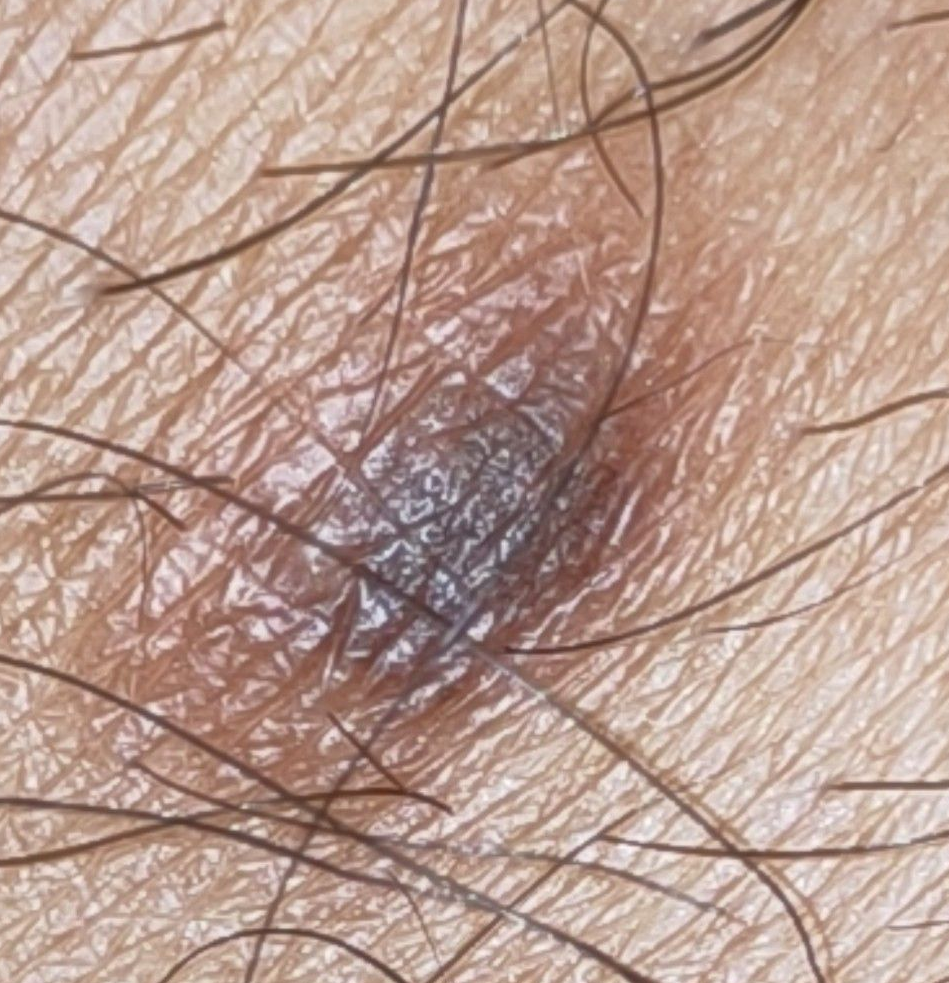
A dermatofibroma is a benign, firm, scar-like growth in the skin. It often appears as a small brown, tan, pink, red-brown, or reddish bump, most often on the legs, and it may dimple inward when pinched. Most dermatofibromas do not need removal. The important decision is whether the bump behaves like a routine dermatofibroma or whether it needs biopsy, complete excision, or referral because melanoma, DFSP, and atypical dermatofibroma variants can mimic a harmless firm bump.
What to remember
The safest mental model is this: a classic dermatofibroma is usually a tiny scar-like bump in the skin, not a dangerous tumor. But the label "dermatofibroma" should be earned by the exam. A changing, bleeding, enlarging, blue-black, facial, recurrent, or unusual lesion deserves a diagnosis-first plan.
Key takeaways
- Dermatofibromas are usually benign, stable, and safe to observe.
- The dimple sign is helpful, but it is not a perfect home test.
- Dermatofibromas are often 3-10 mm and are more common in women, especially on the lower legs.
- The cause is debated: many seem to follow minor trauma, but molecular studies support a benign clonal tumor process in at least some cases.
- Complete excision gives the most reliable diagnostic specimen and the most definitive removal, but the scar is longer than the bump.
- If DFSP is in the differential, superficial shave biopsy is the wrong instinct; tissue depth matters.
Evidence snapshot
- Han and colleagues described 122 dermatofibromas; nearly 80% occurred in patients ages 20-49, and more than 70% were on the extremities ( Han et al., 2011 ).
- Senel and colleagues analyzed 200 dermatofibromas and found a mean age of 42 years, female predominance of 67%, and the leg as the most common location ( Senel et al., 2015 ).
- AAFP describes dermatofibromas as commonly 3-10 mm and notes possible triggers such as minor trauma, insect bites, viral infections, ruptured cysts, or folliculitis ( Shen-Wagner et al., 2024 ).
- Zaballos and colleagues studied 412 dermatofibromas and found the classic central white scar-like patch with peripheral pigment network in 34.7%; Ferrari and colleagues found melanoma-like dermoscopy in 16.2% of 130 lesions ( Zaballos et al., 2008 ; Ferrari et al., 2013 ).
- Cellular dermatofibroma deserves different follow-up when larger than 1 cm and incompletely removed; Gaufin and colleagues found a 10% recurrence rate when margins were involved on initial biopsy ( Gaufin et al., 2019 ).
What is a dermatofibroma?
A dermatofibroma is a benign fibrous growth in the dermis, the deeper layer of the skin. It is also called fibrous histiocytoma or benign fibrous histiocytoma in pathology literature.
Unlike an epidermoid cyst , a dermatofibroma is not a sac filled with keratin. Unlike a lipoma , it is not a soft fatty lump under normal skin. It is a firm, collagen-rich nodule within the skin itself ( Hornick, 2020 ).
Patients usually notice a small firm bump, a brown spot that feels harder than it looks, or a shin lesion that keeps getting nicked while shaving. Many dermatofibromas are painless. Some itch or hurt with direct pressure. Most remain stable for years.
The practical point is not "all firm brown bumps are dermatofibromas." The practical point is: a classic dermatofibroma can often be watched, but a lesion that does not fit the classic story should not be treated casually.
Why did I get a dermatofibroma?
Dermatofibromas have long been explained as localized scar-like reactions after minor skin injury, but newer molecular studies suggest that at least some are true benign clonal growths.
Common possible triggers include a bug bite, shaving nick, folliculitis, ruptured follicle, viral infection, or small trauma the patient may not remember ( Shen-Wagner et al., 2024 ).
That explanation is useful, but incomplete. Immunostaining and molecular work support a mesenchymal and fibroblastic proliferative process, and recurrent protein kinase C gene fusions have been identified in benign fibrous histiocytoma ( Li et al., 1994 ; Plaszczyca et al., 2014 ). In plain language: some dermatofibromas may start after minor skin irritation, but the bump that remains behaves like a real benign growth, not just ordinary scar tissue.
For one or two stable lesions, this debate rarely changes the plan. Multiple eruptive dermatofibromas are different. When many new dermatofibromas appear over a short time, the medical context matters because associations with lupus, HIV, immunosuppressive drugs, and hematologic malignancy have been reported ( Seifi et al., 2022 ).
What does the dimple sign actually prove?
The dimple sign means the center of the bump pulls inward when the surrounding skin is pinched from the sides. It is a useful clue for dermatofibroma, but it is not a perfect diagnostic test.
Dermatologists use the dimple sign because a dermatofibroma is tethered within the dermis, so lateral pressure often makes the center pucker. Meffert and colleagues emphasized that dimpling is not unique to dermatofibroma ( Meffert et al., 1997 ).
Dermoscopy is also helpful, but not absolute. The classic pattern is a central white scar-like patch with a peripheral pigment network; however, dermatofibromas have multiple dermoscopic patterns, including melanoma-like patterns ( Zaballos et al., 2008 ; Ferrari et al., 2013 ).
A surgeon's perspective: In my practice, the dimple sign is a clue, not a permission slip to stop thinking. A small, stable, button-like bump on the shin that dimples beautifully is one category. A lesion that is growing, bleeding, changing color, losing its border, appearing on the face, or simply not fitting the story is another. The safest decision is to match the exam to the behavior of the lesion.
How is a dermatofibroma different from a cyst, lipoma, melanoma, or DFSP?
A dermatofibroma is a firm dermal nodule. A cyst is usually a sac. A lipoma is usually soft, rubbery, and deeper. Melanoma and dermatofibrosarcoma protuberans matter because they can sometimes look deceptively quiet early.
| Feature | Dermatofibroma | Epidermoid cyst | Lipoma | Melanoma | DFSP |
|---|---|---|---|---|---|
| Typical feel | Firm, button-like, fixed in the skin | Round, sometimes with a central punctum | Soft, rubbery, mobile | Variable | Firm plaque or nodule |
| Common color | Brown, tan, pink, red-brown | Skin-colored unless inflamed | Normal overlying skin | Often irregular or multicolored | Pink, red-brown, violet, or skin-colored |
| Common site | Extremities, especially lower legs | Face, trunk, scalp, many sites | Trunk and limbs | Anywhere | Trunk most common, then limbs |
| Dimple sign | Often present | Usually absent | Absent | Usually absent | Usually absent |
| Usual behavior | Stable or slow change | May inflame or drain | Slow enlargement | Progressive change | Slow persistent enlargement over years |
| Biopsy concern | Atypical, changing, facial, symptomatic, large, recurrent, or uncertain | Inflamed, recurrent, unusual, or uncertain | Deep, firm, fixed, painful, fast-growing, or large | Suspicion requires biopsy | Suspicion requires biopsy with adequate depth |
DFSP is not a dermatofibroma that "turned bad." The tumors are molecularly distinct. DFSP is characterized by COL1A1::PDGFB fusion biology and can be misdiagnosed when tissue sampling is too superficial ( NCCN, 2025 ; Zhou et al., 2026 ). DFSP often favors the trunk and shoulder or upper limb region more than the classic lower-leg dermatofibroma. A 2026 JAAD review also notes that Black patients are disproportionately affected, making a high index of suspicion important in skin of color ( Zhou et al., 2026 ).
When should a dermatofibroma be checked?
A suspected dermatofibroma should be checked when the diagnosis is not obvious, the lesion is changing, or the bump is causing symptoms. Observation is reasonable for a classic, stable lesion.
| Schedule evaluation if you notice | Why it matters |
|---|---|
| Rapid growth or steady enlargement | Routine dermatofibromas are usually stable or slow-changing. Growth changes the differential. |
| Bleeding, crusting, or ulceration | Shaving trauma can explain a nick, but spontaneous bleeding deserves evaluation. |
| Irregular color, blue-black color, or new darkening | Pigmented dermatofibroma, aneurysmal dermatofibroma, melanoma, and vascular tumors can overlap clinically. |
| Pain, itch, or increasing tenderness | Symptoms may justify removal and can signal inflammation or another diagnosis. |
| Size greater than 1 cm with cellular features or involved margins | Cellular dermatofibroma may recur when larger or incompletely removed ( Gaufin et al., 2019 ). |
| Size over about 2 cm or indistinct borders | Larger or poorly defined lesions need a more careful diagnostic plan. |
| Facial location | Facial dermatofibromas are uncommon, often clinically misdiagnosed, and recur more often in published series ( Alsawas et al., 2022 ). |
| Multiple new dermatofibromas appearing quickly | Multiple eruptive dermatofibromas can be associated with immune dysregulation or systemic disease ( Seifi et al., 2022 ). |
Which dermatofibroma variants change the plan?
Most dermatofibromas are ordinary benign lesions. Variants matter because some recur more often, mimic melanoma or DFSP, or require a better specimen than a superficial shave can provide.
| Variant or scenario | What to remember |
|---|---|
| Cellular dermatofibroma | Can recur locally, especially when larger than 1 cm and incompletely removed; Gaufin found 10% recurrence with involved margins, lower than older 26-50% reports ( Gaufin et al., 2019 ). |
| Aneurysmal dermatofibroma | Can enlarge quickly and appear blue-black or vascular. In a 2,128-case series, aneurysmal dermatofibroma represented 7.9% of dermatofibromas ( Nishimoto et al., 2024 ). |
| Atypical dermatofibroma | Can mimic more aggressive fibrohistiocytic tumors and may require complete excision and follow-up ( Hornick, 2020 ). |
| Facial dermatofibroma | Rare, roughly 1-2% of dermatofibromas, clinically suspected before biopsy in only 9% in one review, with recurrence rates reported around 14-22% ( Alsawas et al., 2022 ). |
For referring clinicians: what details change management?
The practical question is not "Could this be a dermatofibroma?" The question is whether the clinical behavior, dermoscopy, size, site, and biopsy depth are adequate for the differential.
| Clinical question | Practical provider pearl |
|---|---|
| Dermoscopy | Classic dermatofibroma often shows central white scar-like change and peripheral pigment network, but Ferrari reported melanoma-like patterns in 16.2% of lesions ( Ferrari et al., 2013 ). |
| DFSP dermoscopy | DFSP may show delicate pigment network, vessels, and shiny white streaks; dermoscopy can raise suspicion but does not replace tissue diagnosis ( Bernard et al., 2013 ). |
| Biopsy depth | If DFSP is in the differential, punch, incisional, or core biopsy should include the deeper subcutaneous layer. NCCN specifically warns against inadequate superficial sampling; deep fibrohistiocytic lesions are where clinicopathologic overlap matters most ( NCCN, 2025 ; Zelger et al., 1994 ). |
| Pathology pattern | Dermatofibroma is usually factor XIIIa-positive and CD34-negative; DFSP is usually CD34-positive and relatively factor XIIIa-negative. Exceptions occur, so architecture and depth matter ( Goldblum and Tuthill, 1997 ; West et al., 2014 ). |
| Difficult cases | Stromelysin-3 and D2-40 can support the differential in selected challenging cases, but they complement rather than replace clinicopathologic judgment ( Kim et al., 2007 ; Bandarchi et al., 2010 ). |
Do dermatofibromas need to be removed?
Most dermatofibromas do not need removal. They are benign, often stable, and usually safe to observe when the diagnosis is clear.
Removal is considered when the bump is painful, repeatedly nicked while shaving, cosmetically bothersome, atypical, changing, facial, recurrent, or diagnostically uncertain.
The trade-off is scar honesty. A dermatofibroma is often smaller than the incision needed to remove it. Because the lesion sits in the dermis and can extend downward, definitive excision usually creates a line longer than the original bump. For many patients, a flat planned line is still preferable to a raised, dark, tender nodule. For others, observation is the better cosmetic choice.
A surgeon's perspective: My bias is to be honest about the scar before making the incision. If the diagnosis is classic and the bump is not bothering the patient, observation is often the most elegant choice. If the lesion is atypical or the patient wants it gone, I would rather remove it in a way that gives a clean specimen and a planned scar than chase a tiny opening and leave uncertainty behind.
What are the treatment options?
Treatment depends on the goal. If the goal is diagnosis and definitive removal, full-thickness excision is the most reliable option. If the goal is surface improvement only, partial treatments may be discussed, but they can leave residual firmness or pigment.
| Option | Most appropriate use | Trade-off |
|---|---|---|
| Observation | Classic, stable, asymptomatic dermatofibroma | No scar, but the bump remains. |
| Shave flattening | Selected raised lesions when the diagnosis is secure | Less invasive, but often incomplete because deeper fibrous tissue remains. |
| Cryotherapy or laser-based surface treatment | Selected cosmetic color or surface concerns | May improve appearance but is not definitive removal or diagnostic sampling. |
| Punch biopsy | Small diagnostic sample when partial sampling is appropriate | Can miss architecture and depth; not ideal when melanoma or DFSP is a real concern. |
| Elliptical excision | Symptomatic, recurrently traumatized, atypical, changing, facial, or uncertain lesions | Most reliable diagnostic specimen and most definitive removal, but leaves a longer linear scar. |
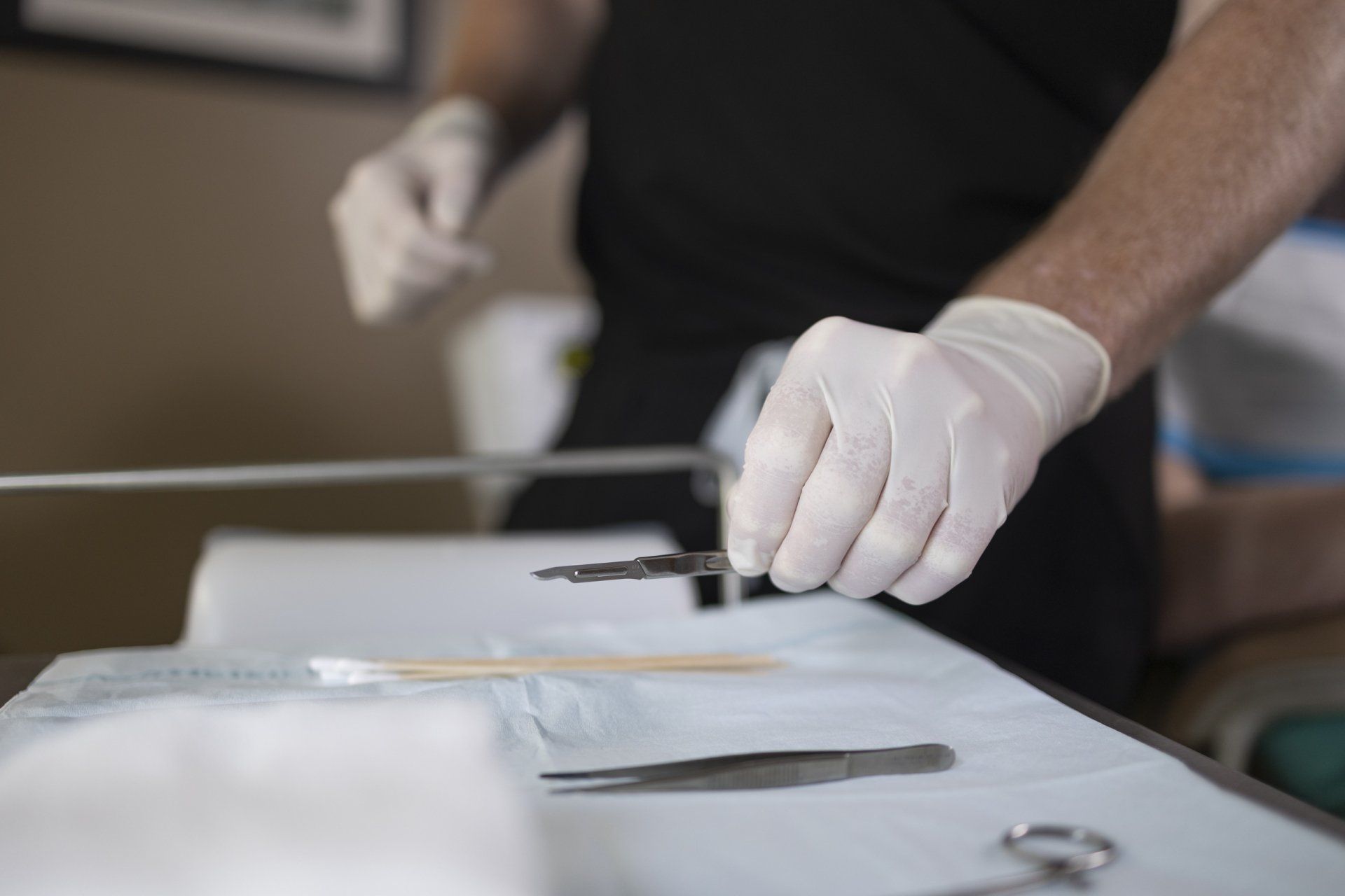
What happens during dermatofibroma excision?
Dermatofibroma excision is usually an in-office procedure under local anesthesia. The skin is numbed, the lesion is removed full-thickness, and the wound is closed in layers to reduce tension.
| Step | Why it matters |
|---|---|
| Exam, marking, and photo when needed | Confirms the target lesion and documents clinical behavior. |
| Local anesthesia | Allows the procedure to be done comfortably in the office. |
| Elliptical full-thickness incision | Gives a complete specimen rather than a surface-only sample. |
| En bloc removal | Preserves architecture for pathology. |
| Layered closure | Reduces tension, especially on the lower leg. |
| Pathology submission | Confirms the diagnosis and screens for atypical variants or mimics. |
Advanced Dermatologic Surgery has an on-site CLIA-certified pathology laboratory used for Mohs surgery and intraoperative frozen-section work when medically necessary. Routine benign excision specimens such as typical dermatofibromas are not the usual purpose of the in-office lab. If an operation reveals something unusual, Dr. Hocker's dermatopathology training and the availability of on-site frozen-section capability allow real-time diagnostic escalation when needed.
What is recovery like after dermatofibroma removal?
Recovery is usually straightforward, but lower-leg scars deserve respect. The shin and lower leg heal more slowly than the face or trunk because of tension, swelling, and less forgiving circulation.
Patients can usually return to light activity quickly, but strenuous exercise, impact, and friction are limited while the wound gains strength.
- Mild soreness and swelling are common for several days.
- A pressure dressing or bandage is usually used at first, followed by simple wound care.
- Compression wrapping or compression stockings may be recommended, especially for lower-leg excisions where swelling can slow healing.
- Surface suture removal is commonly around 10-14 days on the lower leg, depending on tension and location.
- The scar may look pink or darker early, then gradually softens and fades over 12-18 months.
- Scar support may include sun protection and, when appropriate, silicone gel or silicone sheeting after the surface has healed.
Do not stop prescribed blood thinners, aspirin, NSAIDs, fish oil, anticoagulants, or other physician-directed medications unless the prescribing clinician tells you to. Medication decisions should be individualized, especially for patients with heart, stroke, clotting, or vascular history.
How does Advanced Dermatologic Surgery approach dermatofibromas?
Advanced Dermatologic Surgery approaches dermatofibromas by separating three questions: Is the diagnosis secure? Is the lesion bothering the patient? Is the scar trade-off worth it?
That order matters. A patient who wants reassurance may not need surgery. A patient whose shin lesion bleeds every time they shave may benefit from removal. A patient with an enlarging, irregular, facial, blue-black, recurrent, or unusual lesion needs a diagnosis-first plan rather than a cosmetic shortcut.
Dr. Hocker's background in dermatology, dermatopathology, Mohs surgery, and reconstructive dermatologic surgery is most relevant when the lesion is atypical, the scar location is unforgiving, or the procedure needs both diagnostic discipline and careful closure. The goal is not to make a benign bump sound dramatic. The goal is to avoid missing the rare problem while planning the smallest responsible scar.
A surgeon's perspective: I do not treat a changing, bleeding, rapidly enlarging, or clinically odd bump as a cosmetic removal problem. First I protect the diagnosis. Then I plan the scar. That order is what keeps a small office procedure from becoming a missed opportunity to do the right operation the first time.
Advanced Dermatologic Surgery is located at 6901 W 121st Street, Overland Park, KS 66209, serving Overland Park, Kansas City, Leawood, Olathe, Lenexa, Prairie Village, and the broader Kansas City metro. Patients can call (913) 661-1755 or request an appointment online.
Frequently Asked Questions
Is a dermatofibroma skin cancer?
Can a dermatofibroma turn into DFSP?
Why did I get a dermatofibroma?
Are dermatofibromas more common in women?
Will a dermatofibroma go away on its own?
Why does my dermatofibroma get darker after shaving?
Should I biopsy it or remove the whole thing?
Will the scar be bigger than the bump?
Does insurance cover dermatofibroma removal?
When should a primary care clinician refer a suspected dermatofibroma?
When should you schedule an evaluation?
Schedule evaluation if a firm brown bump is new, changing, bleeding, painful, repeatedly irritated, cosmetically bothersome, on the face, larger than expected, recurrent, or not clearly diagnosed.
A short in-person exam can often sort out whether the safest plan is reassurance, dermoscopy, biopsy, or complete excision.
Schedule a Consultation